The Human Anatomy and Physiology Lab Manual 13th Edition is a comprehensive guide designed to simplify complex concepts through clear, step-by-step instructions, suitable for both laboratory and non-laboratory settings, enhancing its versatility and accessibility for students.
Overview of the Lab Manual
The Human Anatomy and Physiology Lab Manual 13th Edition is a user-friendly, comprehensive resource designed to enhance learning through clear, step-by-step guidance. It offers a structured approach to understanding complex anatomical and physiological concepts, with activities and exercises tailored to promote hands-on engagement. The manual is adaptable for both laboratory and non-laboratory environments, making it versatile for diverse learning settings. Its organized format and inclusion of visual aids ensure that students can grasp essential principles effectively, providing a solid foundation for their studies in anatomy and physiology.
Importance of Lab Manuals in Anatomy and Physiology
Lab manuals are essential tools for anatomy and physiology education, providing structured exercises to apply theoretical knowledge. They include dissection guides and practical activities, helping students understand complex anatomical structures. Clear objectives and additional resources enhance learning, making intricate concepts more accessible. These manuals also support visual and kinesthetic learners, offering hands-on experiences that reinforce understanding and retention. As a result, lab manuals are indispensable for developing practical skills and fostering a deeper appreciation of human anatomy and physiology.
Key Features of the 13th Edition
- Updated content with modernized lab exercises for hands-on learning.
- Enhanced visuals, including detailed illustrations and diagrams, to aid comprehension.
- Incorporates digital tools and multimedia resources for a blended learning experience.
- Clear learning objectives and step-by-step instructions for ease of use.
- Improved organization based on user feedback for better accessibility.
These features ensure a comprehensive and engaging learning experience for anatomy and physiology students.
How to Use the Lab Manual Effectively
To maximize the benefits of the Human Anatomy and Physiology Lab Manual 13th Edition, start by reviewing the table of contents to understand the organization. Set dedicated study times to align with lectures or readings. Utilize the clear goals and step-by-step instructions to track your progress. Engage actively by taking notes and completing exercises. Leverage additional resources like dissection guides for visual learning. Study in different settings to enhance flexibility. Break down study sessions into manageable topics to avoid overwhelm. Consistently review and practice to reinforce concepts. Seek help when needed to clarify doubts promptly. Adopting a structured and active learning approach will help you navigate the material confidently and achieve your educational objectives effectively.
Foundational Concepts in Anatomy and Physiology
Grasp the basics of human anatomy and physiology, focusing on the structure-function relationship, levels of organization, and essential terminology to build a strong foundational understanding.
Understanding the Structure and Function of the Human Body
The human body is intricately organized into systems that work together to maintain homeostasis. This section explores the foundational principles of anatomy and physiology, focusing on how structure relates to function. By examining the body’s hierarchical organization, students gain insights into how cells, tissues, organs, and systems interact. Practical exercises and clear explanations simplify complex concepts, enabling learners to grasp essential physiological processes and their significance in overall health. This understanding is crucial for applying anatomical knowledge in real-world scenarios, as emphasized in the lab manual.
Cellular and Tissue Basics
Cells are the fundamental building blocks of life, performing essential functions like metabolism, reproduction, and response to stimuli. Tissues, composed of specialized cells, form structural and functional units of organs. The lab manual explores cellular structure, tissue classification, and their roles in maintaining bodily functions. Histology, the study of tissue structure, and cytology, the study of cells, are emphasized. Practical exercises, including virtual microscopy, help visualize cellular and tissue organization, enhancing understanding of human anatomy and physiology. This section provides a foundation for advanced study of body systems.
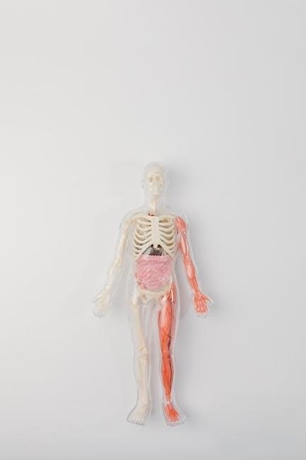
Organization of the Body: Levels of Complexity
The human body is organized into six levels of complexity, starting from the simplest to the most complex: chemical, cellular, tissue, organ, organ system, and organismal. Each level builds on the previous one, forming a hierarchical structure that ensures proper bodily functions. Chemicals form cells, cells form tissues, tissues compose organs, organs make up systems, and systems integrate to create the entire organism. This structured organization allows for specialized functions and efficient communication, enabling the body to maintain homeostasis and respond to external changes effectively.
The Skeletal System
The skeletal system is thoroughly explored, focusing on bones, cartilage, and ligaments, providing detailed insights into their structure, functions, and role in movement and body support, enhanced by clear visuals and practical exercises for hands-on learning.
Classification of Bones
Bones are classified into five categories based on shape and function: long, short, flat, irregular, and sesamoid. Long bones, like the femur, support body weight and enable movement. Short bones, found in the wrist and ankle, provide stability. Flat bones, such as the skull and ribs, protect internal organs. Irregular bones, like vertebrae, have unique shapes for specific functions. Sesamoid bones, such as the patella, reduce friction in joints. This classification aids in understanding their roles in the skeletal system.
Structure and Function of the Axial Skeleton
The axial skeleton comprises the skull, vertebral column, ribs, and sternum, forming the body’s central framework. It provides support, protects vital organs like the brain and heart, and facilitates movement. The skull encases the brain, while the vertebral column shields the spinal cord. Ribs and sternum protect thoracic organs, aiding in breathing. This system ensures structural integrity and flexibility, enabling posture and bodily functions. Its design balances strength and mobility, essential for daily activities and overall bodily stability.
Structure and Function of the Appendicular Skeleton
The appendicular skeleton includes the upper and lower limbs, shoulder and pelvic girdles, enabling movement, support, and protection. It comprises long, short, and irregular bones, with unique structures like the femur and humerus. Its primary functions include facilitating locomotion, providing attachment points for muscles, and supporting the body’s weight. The girdles connect the limbs to the axial skeleton, enhancing stability and range of motion. This system is essential for voluntary movement and maintaining posture, interacting closely with the musculoskeletal and nervous systems to ensure precise and coordinated actions.
Joints and Movement
Joints, or articulations, are points where bones connect, enabling movement and providing stability. The lab manual explores joint classification, including synovial, cartilaginous, and fibrous types, and their functional roles. It details how joints facilitate movements like flexion, extension, and rotation, supported by muscles and ligaments. Practical exercises guide students in identifying joint structures and understanding their biomechanics. Visual aids and activities help clarify how joints contribute to overall mobility and maintain structural integrity, enhancing comprehension of the musculoskeletal system’s dynamic interactions.
The Muscular System
The Muscular System section in the 13th Edition Lab Manual provides a detailed exploration of muscle types, their functions, and physiology, supported by clear instructions for practical understanding.
Types of Muscles and Their Functions
The Human Anatomy and Physiology Lab Manual 13th Edition simplifies the understanding of muscle types, focusing on their roles and locations. Skeletal muscles, attached to bones, enable voluntary movements like walking. Smooth muscles, found in internal organs, function involuntarily, aiding processes like digestion. Cardiac muscle, exclusive to the heart, ensures rhythmic contractions for blood circulation. This organized approach helps learners grasp how each muscle type contributes to bodily functions, enhancing their ability to apply anatomical knowledge in practical scenarios.
Muscle Structure and Physiology
Muscle structure and physiology are essential for understanding movement and bodily functions. Muscles are composed of muscle fibers, each surrounded by a plasma membrane called the sarcolemma. These fibers contain myofibrils, which consist of actin and myosin filaments that slide past each other during contraction. Muscle contraction is initiated by nerve impulses, triggering the release of calcium ions, which facilitate the interaction between actin and myosin. This process requires energy from ATP and is regulated by the nervous system. Different types of contractions, such as isotonic and isometric, serve various functional roles in the body.
The Musculoskeletal System: Interaction Between Bones and Muscles
The musculoskeletal system is a dynamic partnership between bones and muscles, enabling movement and stability. Bones provide structural support, while muscles generate force to move them. This interaction is facilitated by tendons and ligaments, connecting muscles to bones and enhancing flexibility. The lab manual discusses how muscle contractions translate into movement, emphasizing the role of leverage and joint mechanics. Through detailed exercises, students explore how this system adapts to various physical demands, ensuring optimal functionality in diverse activities;
The Nervous System
The nervous system is thoroughly explored in the 13th Edition lab manual, with clear, step-by-step guidance to simplify complex neural concepts for effective learning in any setting.
Neural Tissue and Its Components
Neural tissue forms the structural and functional basis of the nervous system, comprising neurons and neuroglia. Neurons transmit and process information through electrical and chemical signals, while neuroglia provide support, insulation, and protection. The 13th edition emphasizes the roles of astrocytes, oligodendrocytes, and Schwann cells in maintaining neural function and integrity. This tissue’s organization enables rapid communication, essential for controlling bodily functions and responses. The lab manual details these components to enhance understanding of neural tissue’s critical role in human physiology.
Structure and Function of the Central Nervous System
The central nervous system (CNS) comprises the brain and spinal cord, serving as the control center for the body. It processes sensory information, coordinates voluntary movements, and regulates various physiological functions. The brain, divided into cerebrum, cerebellum, and brainstem, handles higher cognitive processes, motor coordination, and autonomic functions. The spinal cord relays messages between the brain and peripheral nerves, facilitating reflex actions. Together, these components ensure seamless communication and integration of bodily systems, enabling adaptive responses to internal and external stimuli.
Structure and Function of the Peripheral Nervous System
The peripheral nervous system (PNS) consists of nerves and ganglia, connecting the central nervous system (CNS) to the body. It is divided into the somatic and autonomic nervous systems. The somatic nervous system controls voluntary movements, while the autonomic nervous system regulates involuntary functions like heart rate and digestion. The PNS transmits sensory information to the CNS and motor signals to muscles and glands, enabling responses to stimuli. Its structure includes nerves, which are bundles of axons, and ganglia, clusters of neurons, facilitating communication between the CNS and body.
- Connects CNS to sensory receptors and effectors.
- Enables both voluntary and involuntary responses.
- Includes nerves and ganglia for signal transmission.
Reflexes and Nerve Pathways
Reflexes are automatic responses to stimuli, involving nerve pathways that transmit signals through sensory neurons, the CNS, and motor neurons. The 13th Edition lab manual provides detailed diagrams and exercises to explore reflex arcs, synapses, and the integration of neural signals. Hands-on activities help students trace nerve pathways and understand how reflexes maintain homeostasis and enable rapid responses. Clear instructions guide learners in identifying key structures and simulating neural transmission, enhancing comprehension of these critical physiological processes.
Circulatory and Respiratory Systems
The lab manual explores blood components, heart structure, and blood vessel functions, alongside respiratory processes like breathing and gas exchange, integrating these systems for optimal bodily function.
Blood and Its Components
Blood is a vital fluid composed of plasma, red blood cells (erythrocytes), white blood cells (leukocytes), and platelets. Plasma, the liquid portion, transports nutrients, hormones, and waste products. Red blood cells carry oxygen throughout the body, while white blood cells play a crucial role in immune defense. Platelets are essential for blood clotting, preventing excessive bleeding. The lab manual explores the structure, function, and clinical significance of blood components, including common disorders like anemia and leukemia, emphasizing their impact on overall health.
The Heart: Structure and Function
The heart is a muscular, hollow organ functioning as the body’s central pump, propelling blood through the circulatory system. It consists of four chambers: the right and left atria, and the right and left ventricles. Blood flows through the heart in a specific pathway, entering via the right atrium, moving to the right ventricle, then to the lungs for oxygenation. Oxygen-rich blood returns to the left atrium, flows to the left ventricle, and is pumped to the rest of the body. The septum separates the heart’s chambers, preventing blood mixing. Valves ensure unidirectional blood flow, maintaining efficient circulation. The heart’s rhythmic contractions are regulated by its intrinsic conduction system, enabling continuous blood circulation essential for cellular oxygenation and nutrient delivery.
Blood Vessels and Circulation
Blood vessels, including arteries, veins, and capillaries, play a vital role in circulating blood throughout the body. Arteries carry oxygenated blood away from the heart, while veins return deoxygenated blood. Capillaries enable nutrient and waste exchange at the cellular level. The lab manual explains blood vessel structure, function, and their role in maintaining blood pressure. Practical exercises and diagrams help students visualize blood flow dynamics and understand circulatory disorders like hypertension and atherosclerosis. This section emphasizes the importance of circulation in overall bodily function and health.
The Respiratory System: Breathing and Gas Exchange
The respiratory system facilitates breathing and gas exchange, essential for oxygenating the blood and removing carbon dioxide. The process involves inhalation, where air enters the lungs through the trachea and bronchi, reaching alveoli for gas exchange. Oxygen diffuses into capillaries, binding to hemoglobin, while carbon dioxide is expelled. The lab manual provides detailed illustrations and exercises to explore lung structure, ventilation mechanisms, and the role of diaphragm and intercostal muscles. Practical activities include measuring breathing rates and analyzing spirometry results to understand respiratory efficiency and its physiological significance.
Digestive System
The Digestive System section in the 13th Edition provides a detailed exploration of the digestive tract, accessory organs, and processes like digestion, absorption, and waste elimination.
Structure and Function of the Digestive Tract
The digestive tract, or gastrointestinal tract, is a tube-like structure responsible for breaking down food into absorbable nutrients. It includes the mouth, esophagus, stomach, small intestine, and large intestine. Each segment specializes in mechanical and chemical digestion. The stomach secretes gastric juices, while the small intestine absorbs nutrients through villi. The large intestine reabsorbs water and stores waste. The digestive tract’s lining, composed of mucosa, submucosa, muscularis, and serosa, facilitates these processes. Peristalsis, regulated by the enteric nervous system, ensures smooth movement of food through the tract, enabling efficient digestion and nutrient absorption.
Accessory Organs of the Digestive System
The accessory organs of the digestive system, such as the liver, pancreas, and gallbladder, play a crucial role in digestion. The liver produces bile, which is stored in the gallbladder and released to emulsify fats. The pancreas secretes digestive enzymes and hormones like insulin and glucagon. These organs work together to break down nutrients, absorb them, and regulate blood sugar levels, ensuring proper digestion and overall bodily function.
Process of Digestion and Absorption
Digestion begins with ingestion, where food is broken down mechanically in the mouth and chemically by enzymes. The stomach further processes food into a liquid mixture, which enters the small intestine for nutrient absorption; Specialized enzymes break down carbohydrates, proteins, and fats into simpler forms like glucose, amino acids, and fatty acids. These nutrients are absorbed into the bloodstream through intestinal lining cells, facilitated by finger-like projections called villi. The bloodstream and lymphatic system transport these nutrients to cells throughout the body for energy and repair, ensuring proper bodily functions and overall health.
Common Disorders of the Digestive System
Common digestive system disorders include gastroesophageal reflux disease (GERD), irritable bowel syndrome (IBS), inflammatory bowel disease (IBD), and peptic ulcers. These conditions often result from factors like poor diet, stress, or infections. Symptoms may include abdominal pain, bloating, diarrhea, or constipation. Diagnoses typically involve endoscopic exams or imaging tests. Treatments range from dietary changes and medications to surgery in severe cases. Understanding these disorders is crucial for maintaining digestive health and preventing complications. Early detection and proper management can significantly improve quality of life for individuals affected by these conditions.
Endocrine and Urinary Systems
The Endocrine and Urinary Systems section explores hormone regulation, kidney function, and fluid balance, with practical exercises to enhance learning and understanding of these processes.
Overview of the Endocrine System
The endocrine system is a network of glands that produce and secrete hormones, regulating various bodily functions such as metabolism, growth, and reproductive processes. Key endocrine organs include the pituitary gland, pancreas, thyroid, adrenal glands, and reproductive organs like the ovaries and testes. These hormones help maintain homeostasis, influencing mood, energy levels, and overall health. The endocrine system works closely with the nervous system to ensure proper bodily functions, making it essential for maintaining health and well-being.
Major Endocrine Glands and Their Hormones
The major endocrine glands include the pituitary, thyroid, adrenal, pancreas, and gonads (ovaries and testes). The pituitary gland regulates other endocrine glands. The thyroid produces hormones like thyroxine, controlling metabolism. The adrenal glands release adrenaline and cortisol, managing stress and electrolyte balance. The pancreas secretes insulin and glucagon, regulating blood sugar. The gonads produce sex hormones, such as estrogen and testosterone, essential for reproductive processes. These glands work harmoniously to maintain homeostasis and overall bodily functions.
Structure and Function of the Urinary System
The urinary system, also known as the renal system, consists of the kidneys, ureters, bladder, and urethra. Its primary function is to remove waste and excess fluids from the body by producing urine. The kidneys filter blood to eliminate toxins, regulate electrolytes, and maintain pH balance. The ureters transport urine to the bladder for storage. The urethra then releases urine during urination. This system plays a crucial role in maintaining homeostasis and overall health, as detailed in the lab manual.
Regulation of Water, Electrolytes, and pH
The regulation of water, electrolytes, and pH is crucial for maintaining homeostasis. The kidneys play a central role by filtering blood, removing excess substances, and adjusting fluid balance. Hormones like aldosterone and antidiuretic hormone (ADH) help regulate electrolytes and water retention. The lungs contribute to pH balance by expelling CO2. This process ensures proper cellular function and overall health. The lab manual provides detailed exercises to explore these mechanisms, emphasizing the importance of understanding how the body maintains equilibrium in dynamic conditions.
Reproductive System
The Reproductive System section in the 13th Edition lab manual provides detailed insights into male and female anatomy, fertilization processes, and common reproductive health issues, supported by clear illustrations.
Structure and Function of the Male Reproductive System
The male reproductive system includes the testes, penis, and accessory glands like the epididymis, vas deferens, seminal vesicles, and prostate gland. The testes produce sperm and testosterone, while the penis facilitates sexual intercourse and urination. The epididymis stores sperm, and the vas deferens transports it during ejaculation. Seminal vesicles and the prostate gland secrete fluids that support sperm viability. Disorders such as erectile dysfunction or infertility can affect system function, emphasizing the importance of understanding its anatomy and physiology for diagnostic and therapeutic purposes.
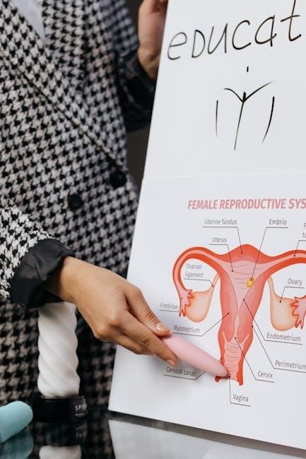
Structure and Function of the Female Reproductive System
The female reproductive system is a complex network of organs and tissues designed for reproduction. It includes the ovaries, which produce eggs and hormones like estrogen and progesterone, the fallopian tubes for fertilization, the uterus to support fetal development, and the cervix and vagina for childbirth and menstrual flow. These components work together to facilitate conception, pregnancy, and childbirth, while also maintaining hormonal balance essential for overall health and reproductive function.
Process of Fertilization and Embryonic Development
Fertilization begins with the fusion of a sperm and an egg, forming a zygote. This process typically occurs in the fallopian tube, initiating embryonic development. The zygote undergoes cleavage, dividing into a morula, and later a blastocyst, which implants in the uterine lining. Gastrulation follows, establishing the three primary germ layers: ectoderm, mesoderm, and endoderm. These layers differentiate into specialized tissues and organs, guided by genetic and environmental factors. Hormonal regulation ensures proper development, leading to the formation of a viable embryo ready for further growth and maturation.
Reproductive Health and Disorders
This section explores common reproductive health issues and disorders, such as sexually transmitted infections, infertility, and hormonal imbalances. It provides clear learning goals, step-by-step instructions, and practical exercises to enhance understanding. The lab manual includes detailed diagrams and real-world examples to illustrate conditions like endometriosis, prostate disorders, and menstrual irregularities. Practical activities focus on diagnostic techniques and prevention strategies, ensuring a comprehensive approach to studying reproductive health and its challenges. This makes the content accessible and engaging for students.

Practical Applications and Resources
The lab manual offers practical exercises, dissection guides, and troubleshooting tips, enhancing hands-on learning and providing additional study tools for mastering human anatomy and physiology effectively.
Lab Exercises and Activities
The 13th Edition lab manual offers a wide range of engaging exercises and activities designed to reinforce anatomical and physiological concepts. These include hands-on experiments, such as histology slide examinations, dissection exercises, and physiological measurements. Interactive simulations and case studies provide real-world applications, while group activities and lab reports encourage collaborative learning. The manual also features assessment tools to track progress, ensuring a comprehensive understanding of human anatomy and physiology. Each activity is tailored to enhance practical skills and critical thinking, making complex topics accessible and engaging for students.
Using Dissection Guides for Better Understanding
Dissection guides in the 13th Edition lab manual offer detailed, step-by-step instructions to enhance hands-on learning. They provide clear visuals and practical tips, making complex anatomical structures easier to comprehend. By following these guides, students can master dissection techniques, fostering a deeper understanding of human anatomy. The inclusion of visual aids and real-world applications ensures that learners can connect theoretical knowledge with practical skills, making the dissection process both educational and engaging.
Additional Study Resources and Tools
Supplement your learning with the 13th Edition lab manual by utilizing online platforms offering interactive simulations, 3D models, and virtual dissections. Access digital flashcards, video tutorials, and practice quizzes to reinforce concepts. The manual also provides links to anatomical atlases and study guides, enhancing your understanding of complex structures. Mobile apps like anatomy labeling tools and physiology simulators further support self-study. These resources ensure a well-rounded learning experience, catering to diverse learning styles and preferences for mastering human anatomy and physiology effectively.
Troubleshooting Common Challenges in Lab Work
Lab work in anatomy and physiology can present challenges, such as difficulty in identifying structures or interpreting data. The 13th Edition lab manual offers practical solutions, including clear instructions, visual aids, and troubleshooting tips. It addresses common issues like specimen handling and equipment usage, ensuring students can overcome obstacles effectively. The guide also provides step-by-step approaches to complex procedures, enhancing understanding and practical skills. By addressing these challenges, the manual serves as an invaluable resource for successful lab experiences.